Of brains and mummichogs: David Hollis leads team in quest to understand how fish brains self-repair
Here’s what biologists know – the brain of mammals is woefully limited in its ability to repair itself. In the face of traumatic brain injury, there is precious little, if any, repair with regard to regenerating new cells to replace the damaged ones.
Fish, on the other hand, have an amazing ability to bounce back after brain injury. Not only do fish have the ability to regrow new brain cells, they are able to construct favorable molecular conditions in which neural tissue can self-repair.

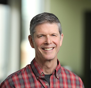
Biology Professor David Hollis
Furman Professor of Biology David Hollis and five students – biology graduates Ellie Bisese ’20, Chandler Ciuba ’20 and Alanksha Kaushik ’19, and neuroscience graduate Amelia Davidson ’20 and Sabrina Mullen, a senior neuroscience major, – collaborated with scientists at the Medical University of South Carolina (MUSC) and Queens University in Belfast (QUB), Northern Ireland to learn more about the superpowers of a particular species of fish. Their findings were published in Molecular Brain.
The researchers set out to provide a profile of the whole genome response of the adult brain to injury in a vertebrate that has the ability to successfully repair neural tissue. Using strict protocol for the use of animals in the lab, the team looked at the response of the mummichog (Fundulus heteroclitus), a diminutive Atlantic Coast fish that inhabits brackish waters like those in estuaries and salt marshes.
Hollis says as vertebrates, fish and mammals share many of the same genes, but the differences in how the two respond to brain injury are striking.
“Mammals respond with various cellular impediments to recovery, whereas, fish respond with a distinct series of cellular events that lead to recovery,” he said.
Hollis said that cellular events are dictated by patterns in how genes are expressed. So, for Hollis and his team, a fundamental question arose: “How do the patterns of gene expression in the injured fish brain compare to what’s known to occur in mammals?”
Bisese of Richmond, Virginia, said the team set its sights on confirming previous findings.

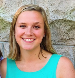
Ellie Bisese ’20
“In science, you have to constantly re-check results because you can never take the first finding at face value,” she said. “Our project focused on identifying what genes are upregulated one hour post-injury. We had basically already found these genes through our previous work with MUSC and Queens, and our work at Furman sought to validate these genes.”
In terms of human application, Hollis said it’s possible that the patterns of gene expression in the recovering brain tissue of fish tell us what genes may be intrinsic to successful neural repair.
“And from this,” he said, “the sources of recovery limitation in the human brain can be further deduced, uncovering potential therapeutic possibilities.”
Apart from the knowledge gained about neural repair, Bisese says the study boosted her confidence.
“Going into the project, I didn’t trust my skills as a scientist,” she said. “But two days into it, I realized I was more capable in the lab than I thought.”.
With renewed confidence in her lab smarts, Bisese plans to take a couple of gap years working in a science or health care-related field before enrolling in medical school. “My dream is to work in obstetrics and gynecology,” she said.
As for future research projects, Hollis says this round of research lays the groundwork for examining new questions.
He said many of the genes they identified have never been implicated in the process of brain repair and regeneration, despite their known functions in relevant cellular events, such as their influence on the proliferation of new cells.
“So, a logical question becomes, ‘What are the proteins for which these genes encode and what are their functions in the reparative process, and how do we test their functional relevance’?” he said. “We can also ask, ‘Where exactly in the brain is this particular gene having its sphere of influence during the recovery process?’”
This latest project, funded by a grant from the South Carolina IDeA Networks of Biomedical Research Excellence, allowed students at a primarily undergraduate university to engage in collaborative efforts with labs at larger research universities.
“This kind of collaboration is reflective of how much of science is done in the modern era,” he said.
Hollis also underscores the inherent value students derive from being involved in faculty research.
“Faculty and student collaboration is an obvious way to give students a unique learning experience that is simply unattainable in a formal classroom,” he said. “It’s rewarding to see how student self-reliance improves as their work progresses in the research setting. And for some, the experience often plays a key role in shaping their decisions regarding their long-term career paths.”